One Atlas from Many Complex Pieces: siibra Brings Scattered Brain Data Together
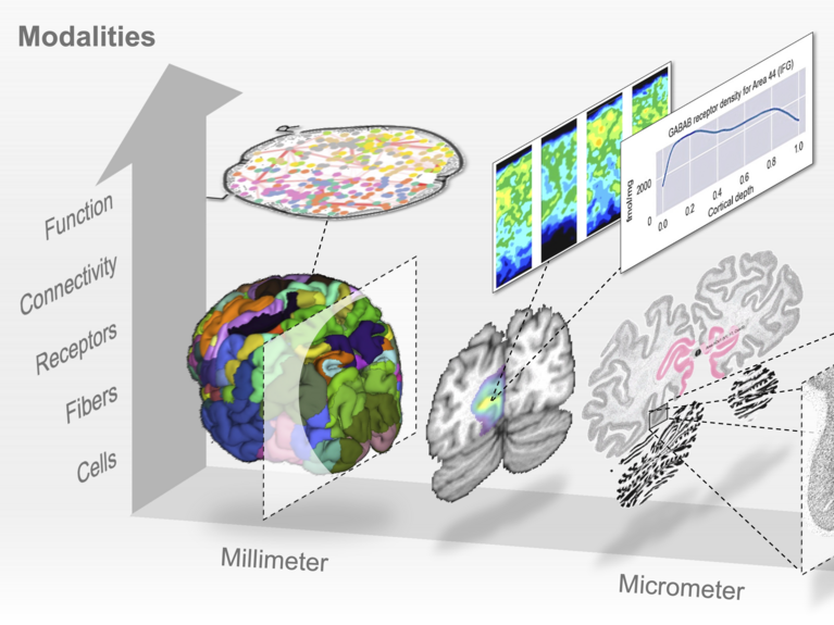
The brain atlas developed with siibra connects brain regions and the variability of brain structure with multiple levels of brain organization, from cells to large-scale networks. (Forschungszentrum Jülich)
Anyone trying to study the human brain runs into the same problem: the evidence lives in different places, at different scales, and speaks different technical languages. A microscopy slide showing individual cells, an MRI scan mapping large-scale anatomy, and a connectivity dataset tracing fiber pathways are all, in principle, describing the same object, but they rarely line up cleanly.
siibra ("Software Interfaces for Interacting with Brain Atlases") is built to solve that. The paper announcing it appears this week in Nature Methods, with first author Timo Dickscheid, Helmholtz AI Associate and member of the Helmholtz AI Steering Board, co-author Christian Schiffer, Helmholtz AI Principal Investigator, and last author Katrin Amunts (Director of INM-1), Helmholtz AI associate, alongside an international research team anchored at the Institute of Neuroscience and Medicine (INM-1), Forschungszentrum Jülich.
Through an interactive 3D viewer, a Python library, and a web API, siibra brings distributed brain data into one shared reference frame and hands it back out in a form other tools can use, whether that's a researcher exploring the atlas visually or a pipeline running fully automated, reproducible analysis. Inside EBRAINS, Europe's neuroscience research infrastructure, this is what makes the Human Brain Atlas work day to day: it's the layer that lets a coarse whole-brain space like MNI communicate with a microscopic model like BigBrain, with the cytoarchitectonic maps of Julich Brain sitting at the anatomical core of that connection.

The siibra tool suite integrates data from diverse sources into a comprehensive atlas of the human brain, making information on brain architecture usable for neuroscience, medicine, and AI development — from the molecular and cellular to the macroscopic level.
High-resolution 3D maps of brain areas and quantitative measurements of brain organisation included in siibra were computed using recent AI approaches for scientific image analysis developed by the Helmholtz AI unit in Jülich. This includes, amongst others, foundation models for microscopic images of cellular brain structure developed by Christian Schiffer et al. (CytoNet), and cell instance segmentation models by Eric Upschulte et al. (Contour Proposal Networks; CPN).
The atlas doesn't force data into rigid boundaries, either. Because no two brains are anatomically identical, siibra works probabilistically: a given coordinate, region, or image patch is treated as belonging to a brain area with some degree of confidence, rather than being forced across a fixed line. That matters when aligning imaging, histology, or electrophysiology data against the same anatomical reference. It also handles genuinely large files without choking on them, meaning a gigabyte- or terabyte-sized imaging dataset can be pulled apart and queried one region at a time, with no need to download the whole thing first.
One test case in the paper concerns deep brain stimulation. Reanalyzing previously published data through siibra, the team finds hints that the VIM, an important relay structure in the thalamus, may sometimes get confused with a neighboring region when clinicians are choosing where to stimulate. Tying detailed microstructural maps to a patient's own scans, the authors argue, could make that kind of targeting more precise.

Dr. Timo Dickscheid, © Forschungszentrum Jülich / Ralf-Uwe Limbach
“For several years, my colleagues and I have been working to turn the vision of a multilevel human brain atlas into a practical resource for neuroscience. While many researchers already use it through the EBRAINS Human Brain Atlas, this publication finally captures the conceptual and technical foundations behind it. For me, it marks an important milestone, documenting years of development and refinement that have culminated in a resource now used by a growing international research community.”
- Timo Dickscheid, Forschungszentrum Jülich
Where the research goes next depends largely on how much additional high-quality brain data becomes available, and how efficiently it can be accessed globally. The team plans to keep building on siibra with more AI-driven analysis tools, and links to further international data platforms are already on the roadmap.
Beyond the atlas itself, siibra's more lasting contribution may be structural: turning specialist-only, terabyte-scale brain data into something any research group can query and build on, rather than only the labs with the infrastructure to handle it alone.
Original publication: Dickscheid, T., Gui, X., Simsek, A. N., Schiffer, C., Mangin, J.-F., Leprince, Y., Jirsa, V., Bjaalie, J. G., Leergaard, T. B., Bludau, S., & Amunts, K. (2026). Siibra: A software tool suite for realizing a Multilevel Human Brain Atlas from complex data resources. Nature Methods. doi: 10.1038/s41592-026-03159-x
- Software: https://siibra.eu
- Article: https://www.nature.com/articles/s41592-026-03159-x
- EBRAINS: https://ebrains.eu